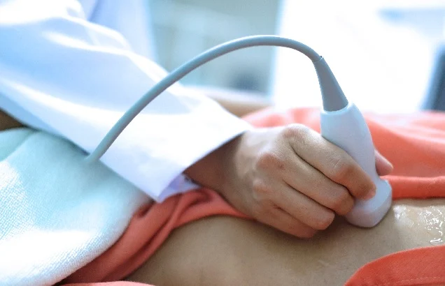
An echocardiogram, also called an echo, is an ultrasound examination of the heart’s anatomy and function. Port Saint Lucie echocardiogram can detect various problems such as cardiomyopathy and valve dysfunction. It is also a non-invasive technique that creates no radiation and has few adverse effects. There are several types of echo tests, such as;
- Transthoracic echocardiogram
The transthoracic echocardiogram is the most prevalent type of echocardiogram test. This exam involves positioning an ultrasound wand known as a transducer on the outside of your chest, near the heart. The device then transmits sound waves through your chest and into the heart.
Additionally, applying gel to your chest aids the sound waves to travel better. These waves bounce off your heart and produce images of your heart structures on a screen.
- Transesophageal echocardiogram
A transesophageal echocardiogram employs a smaller transducer attached to the end of a long tube. The patient will swallow the tube to insert it into the esophagus, which runs behind the heart and joins the mouth and stomach.
Since it provides a “close up” view of the heart, this kind of echocardiogram produces more detailed images of the organ than the conventional transthoracic echocardiogram.
- Doppler ultrasound
Doctors use doppler ultrasounds to examine the flow of blood. They produce sound waves at specified frequencies and observe how they bounce off and return to the transducer. Also, colored doppler ultrasounds can be used by specialists to map the direction and velocity of blood flow in the heart.
Blood flowing toward the transducer shows red, while blood flowing away appears blue. A doppler ultrasound can detect abnormalities with valves or holes in the heart’s walls and analyze how blood flows through it.
- Fetal echocardiogram
A fetal echocardiogram is performed on expectant mothers between weeks 18 and 22 of pregnancy. The transducer is placed across the pregnant woman’s abdomen to check for fetal cardiac abnormalities. The test is considered safe for an unborn child since it does not utilize radiation, unlike an X-ray.
- Stress echocardiogram
A clinician might order an echocardiogram as part of a stress test. A stress test involves physical exercise, like walking or jogging on a treadmill. During the test, your specialist will monitor heart rate, blood pressure, and the heart’s electrical activity.
Moreover, a sonographer will take a transthoracic echocardiogram after and before the exercise. Doctors utilize stress tests to identify ischemic and coronary heart disease, heart failure, or abnormalities with the heart valves.
- Three-dimensional echocardiography
A three-dimensional (3-D) echocardiogram utilizes either transesophageal or transthoracic echocardiogram to generate a 3-D image of your heart. This involves numerous pictures from various angles. It is used before heart valve surgery and to diagnose heart issues in kids.
An echocardiogram is a necessary test that might reveal a lot about your heart’s structure and function. If your clinician suggests an echo, ask about what kind you will be receiving and what to expect. You may require more than one echo or several tests with different techniques to give your physician enough information about your heart.
Additionally, ask your specialist to explain the images to you and assist you in understanding what they mean. Taking an active role in your care and diagnosis might help you feel at ease throughout the process. Call TLC Medical Group Inc or book your consultation online to find out which echocardiography test is best for you.

